Cerebral circulation, cerebral circulation anatomy, venous circulation of the brain & CSF
The venous drainage of the cerebrum happens through two groups of venous blood vessels - the superficial and deep cerebral veins. Check it out. cerebral hemispheres. Lobes, gyri and sulci. Cerebral blood vessels. Brainstem, cerebellum, cerebral hemispheres. Lobes, gyri and sulci. Cerebral blood vessels. Anatomy.app. 3D Anatomy Media Library;

Dural Venous Sinuses Craniosacral therapy, Medical anatomy, Neurology
- This video describes the anatomy of the venous drainage of the brain (including MCQ, practical questions and interactive questions).
Venous Drainage of the CNS Cerebrum TeachMeAnatomy
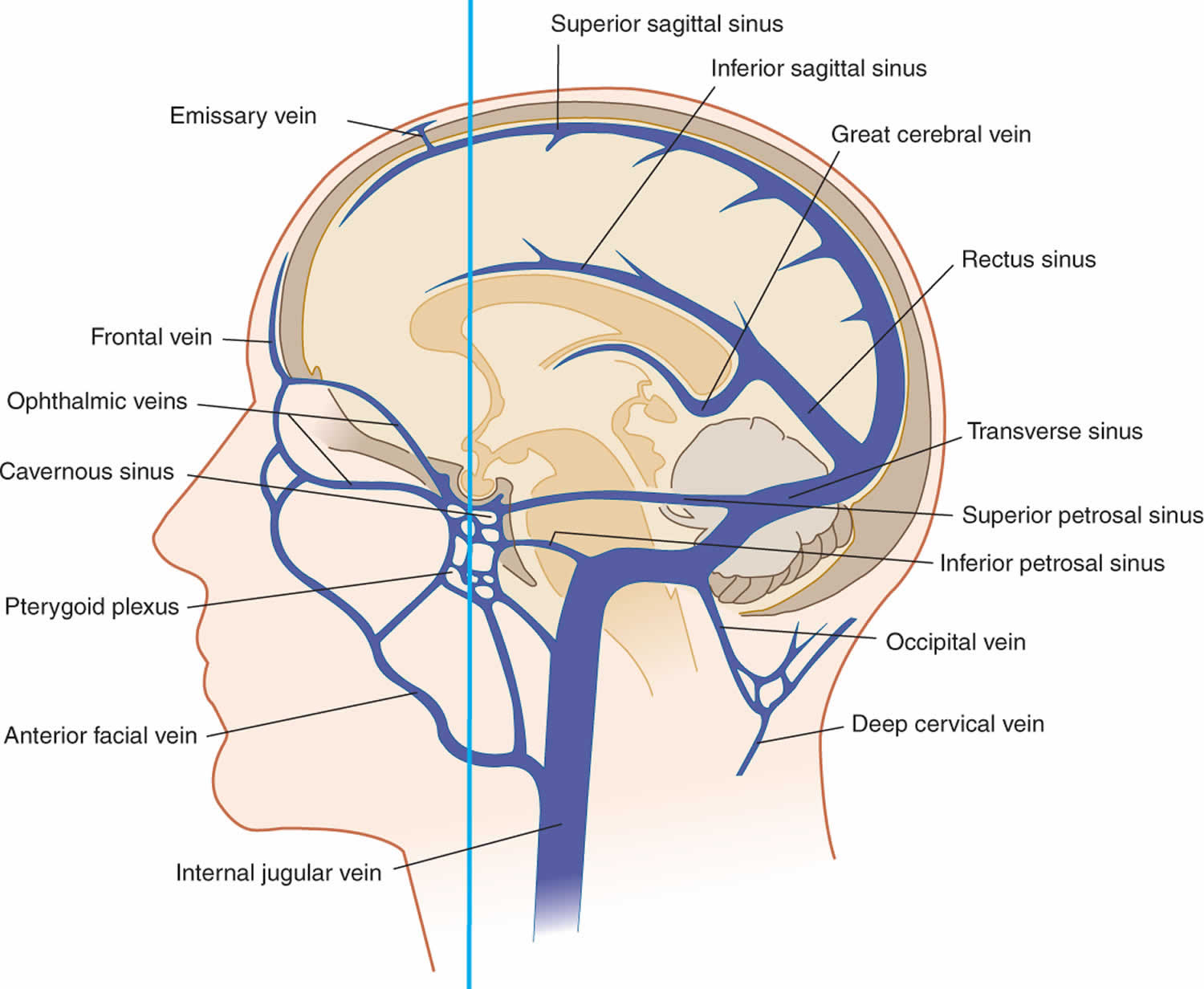
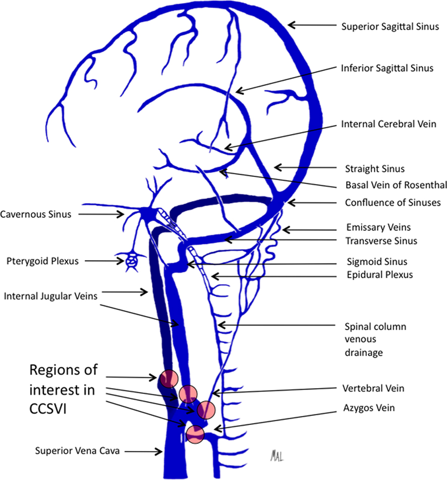
Venous drainage of the brain occurs through a system of cerebral and cerebellar veins, which in turn drain into the dural venous sinuses. The dural venous sinuses ultimately empty into the internal jugular veins which, together with the external jugular vein (draining the scalp and face), returns venous blood from the head and neck region back.
Venous Drainage of the Brain Earth's Lab
The venous drainage of the cerebrum can be separated into two subdivisions: superficial and deep.. Cerebral blood flow (CBF) is the blood supply to the brain in a given period of time. In an adult, CBF is typically 750 millilitres per minute or 15.8 ± 5.7% of the cardiac output.

venous drainage of the brain, inferior view Diagram Quizlet
Venous Drainage of the Brain , Raimund Kleiser & Peter Strasser Chapter First Online: 13 December 2019 3298 Accesses Abstract The venous blood is collected by the veins, drained into the dural sinuses and emptied into the jugular vein by emissary veins, cerebral veins, dural sinuses, internal jugular vein, and brachicephalic vein.

Pin on NEURO
The Venous Drainage of the Central Nervous System star star star star star_half based on 77 ratings Original Author (s): Sam Barnes Last updated: July 18, 2023 Revisions: 43 format_list_bulleted Contents add The central nervous system consists of the cerebrum, cerebellum, brainstem and spinal cord.

Venous Drainage of The Brain Learn Human Anatomy
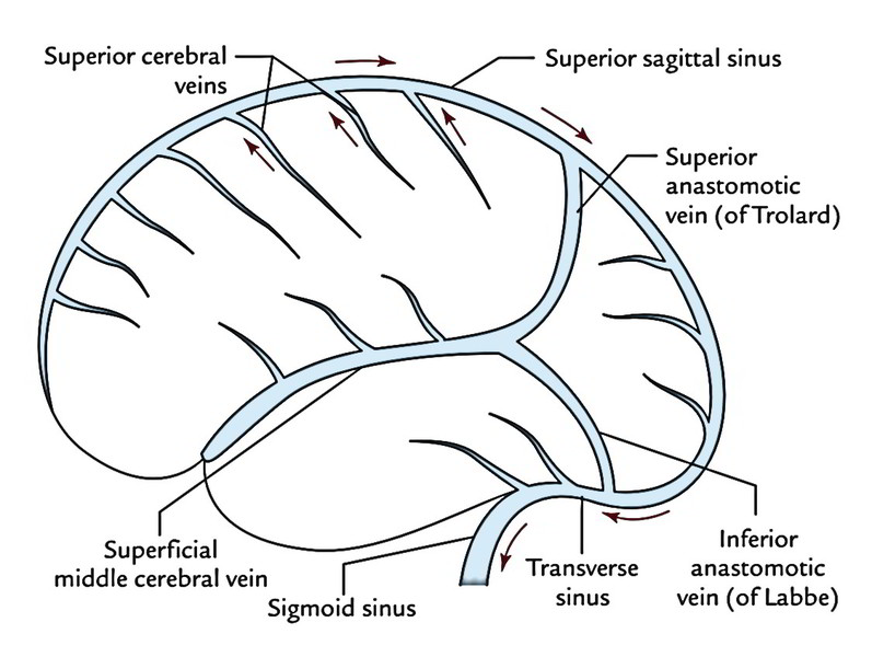
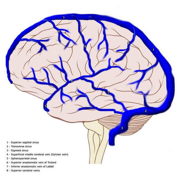
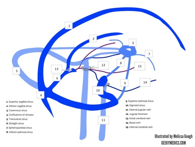
The cerebral veins drain the brain parenchyma and are located in the subarachnoid space. They pierce the meninges and drain further into the cranial venous sinuses. The cerebral veins lack muscular tissue and valves. The cerebral venous system can be divided into: superficial (cortical) cerebral veins deep (subependymal) cerebral veins

Cerebral venous thrombosis state of the art diagnosis and management Semantic Scholar
Delicate venous drainage from the cerebral hemispheres emerges from the brain to form small venous structures in the pia mater. These larger venous channels then form cerebral veins, which bridge the subarachnoid space and enter into endothelial-lined sinuses within the dura mater.
Dural sinuses and encephalic veins anatomy [70]. Download Scientific Diagram
Venous drainage of the brain and meninges: Supplied by the dural venous sinuses. Venous drainage of the scalp and face: Drained by veins synonymous with the arteries of the face and scalp. These empty into the internal and external jugular veins. Venous drainage of the neck: Carried out by the anterior jugular veins.
Venous Drenage Of Brain Kypho
Venous Drainage of the Brain. VI. Neurons as Cells. Neurons and Glial Cells. Techniques Used to Study Neurons at the Cellular Level. Neuronal Shape and Neuronal Information Processing. Myelination. Organelles. Energy Production. Energy Allocation. Glutamate Management. The Endosomal System.

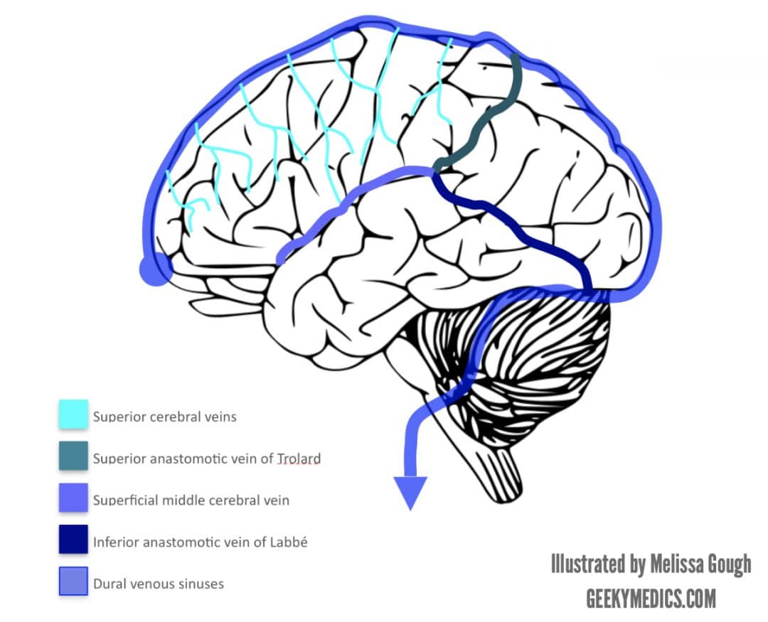
Venous Drainage of the Brain Anatomy Geeky Medics
Cerebrum Veins draining the brain parenchyma may be divided into superficial and deep veins. The superficial veins primarily drain the cerebral cortex, whereas the deep veins drain the deep structures within the hemispheres.
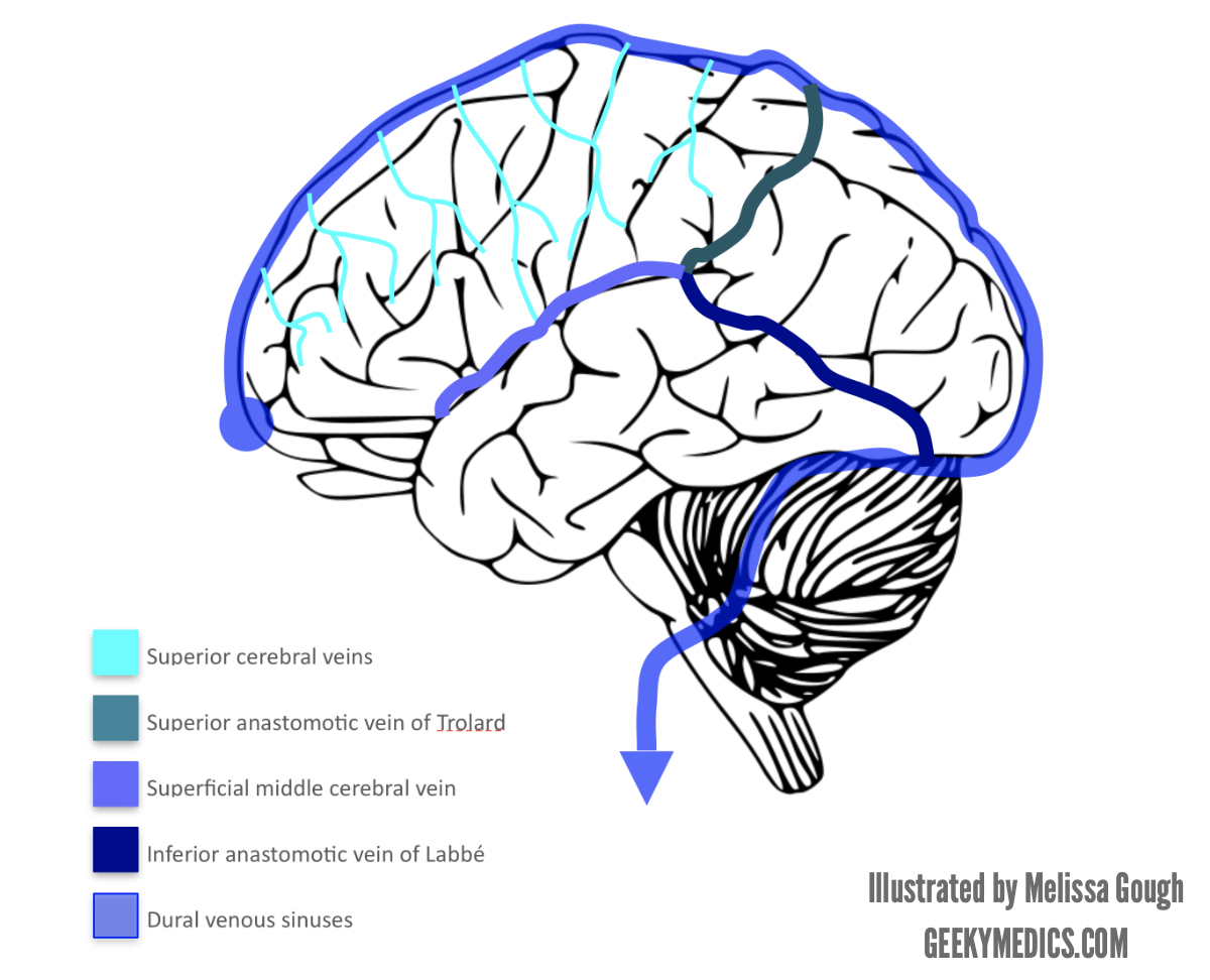
Venous Drainage of the Brain Anatomy Geeky Medics
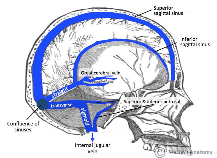
The venous drainage of the brain does not follow the arteries of the brain. Instead, they drain to the dural sinuses, which subsequently drain to the internal jugular vein. Generally, the walls of these drainage pathways are formed by visceral periosteum and dural reflection, both lined with endothelium. The inferior sagittal and straight.
myneurologytips Cortical Venous System
They drain the brain, eyes, meninges, and part of the face through the pterygoid plexus. Additionally, the dural venous sinuses drain the cerebrospinal fluid through arachnoid granulations and allow cerebrospinal fluid to return to the bloodstream. [4] Unlike other veins in the body, the cerebral veins have no muscular walls or valves.

Emergency Medicine EducationCerebral Venous Thrombosis Pearls and Pitfalls
All venous drainage occurs through dural venous sinuses that drain toward the neck veins. The walls of dural venous sinuses are also home to meningeal lymphatic vessels ( 7, 8 ), with a role in the drainage of CSF.
Venous Drainage of the Brain Anatomy Geeky Medics
This video provides a walkthrough of the venous drainage of the brain, including the superficial veins of the cerebral cortex and the dural venous sinuses. Y.
Drenaje venoso del cerebro Anatomía My Star Idea
This chapter describes the vascular anatomy of the brain including the arterial supply and venous drainage. It begins by describing the anterior circulation forming from the carotid arteries, common variants, and fetal remnants. The anterior circulation has several anastomoses to posterior circulation. The posterior circulation supply emanates.